SVETLANA PANASYUK
Medical Hyperspectral Imaging
Optical Metrology
Tissue Spectroscopy
Mantle Flow
GPS
Remote Sensing
Image Processing
Fun
Reference Earth Model
|
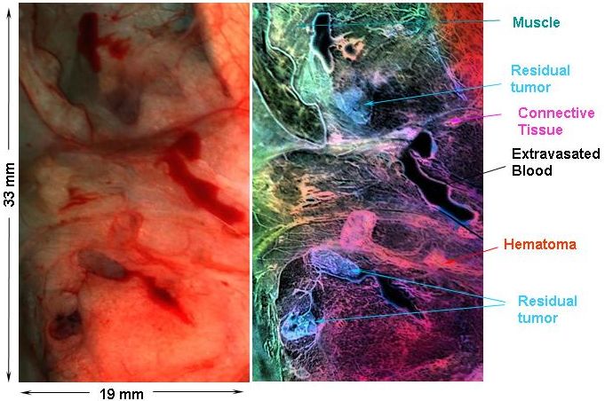
Hyperspectral imaging consists of acquisition and analysis of images
recorded using a number of different wavelengths of light (spectral bands).
If we capture the same object on many bands of the spectrum, we could
generate a data cube - each slice revealing different information
depending on the chemical properties of the object. Different types of tissue have different composition, physical and chemical. Therefore, once an algorithm is created to distinguish each tissue type, the data could be presented in such a way that facilitates a surgeon during an operation. Here is an example of an image taken during an open surgery and processed later to identify areas of residual tumors (cyan color):
Work was done while at HyperMed, Inc: HyperMed |
 Hyperspectral Imaging Cancer Application